What is a PE (pulmonary embolus)?
Working from the earlier post, when a blood clot in the vein breaks off and travels north, it passes through the heart and into the blood vessels (pulmonary arteries) that carry blood to the lungs to offload CO2 and take up Oxygen to then deliver back out to the body. The reasons PEs cause issues is two fold. One, large enough clots can actually block enough outflow from the heart to put significant strain on the heart even causing cardiac collapse. Two, they create areas in the lungs which are being ventilated (trying to exchange CO2/O2) as you breathe but not being perfused (no blood flow therefore, no gas exchange). If large enough segments of the lungs are affected cardiopulmonary collapse can occur. Shortness of breath and chest pain is a common presenting symptom.
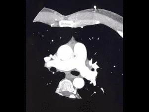
Below is a CT angio done to assess the heart arteries. The bright white tubes in the middle of the picture is the aorta (11 and 5 o’clock) and the pulmonary artery which branches left and right to go to each lung. They are bright white because of IV contrast. As you follow the vessels out into the lungs (the large basically empty spaces on either side of midline) you can see the arteries getting smaller and smaller. Some are imaged in cross section so they appear as white dots. The heart is the large central mass seen as you scan down. The artery is white because of contrast given intravenously. In the smaller vessels you can see areas of gray within the white. These are the areas where thrombus has been lodged.